bise Rawalpindi board biology notes for grade 9 Chapter 9 Transport Pdf transport, Important short question, Understanding The Concepts, Science, Technology And Society, and Mcqs Pdf Download Now.
Mcqs on Transport G9
i) In most plants, food is transported in the form of;
- A. Glucose
- B. Sucrose
- C. Starch
- D. Proteins
ii) Stomata close when guarding cells;
- A. Lose water
- B. Gain chloride ions
- C. Become turgid
- D. Gain potassium ions
iii) Trace the pathway of water from the soil through the plant to the atmosphere.
- A. Endodermis, cortex, epidermis, xylem, intercellular spaces in mesophyll, stomata
- B. Epidermis, endodermis, phloem, cortex of leaf, intercellular spaces of mesophyll, stomata
- C. Root hairs, epidermis, cortex, xylem, endodermis, intercellular spaces in mesophyll, stomata
- D. Root hairs, cortex, endodermis, xylem, intercellular spaces in mesophyll, stomata
iv) When fibrinogen makes blood clot it separates from blood and the remainder is called;
- A. Plasma
- B. Lymph
- C. Serum
- D. Puss
v) What is correct about human red blood cells?
- A. Have limited life span
- B. Are capable of phagocytosis
- C. Produce antibodies
- D. Are multinucleate
vi) Which of the following tissue layer is found in all blood vessels?
- A. Smooth muscle
- B. Endothelium
- C. Skeletal muscle
- D. Connective tissue
vii) When does the atria contract?
- A. Before diastole
- B. After systole
- C. During diastole
- D. During systole
viii) Which of the following contains deoxygenated blood in an adult human?
- A. Left atrium
- B. Pulmonary artery
- C. Pulmonary vein
- D. All of the above
ix) Which of the following chambers has the thickest walls in the human heart?
- A. Right atrium
- B. Left atrium
- C. Left ventricle
- D. Right ventricle
x) Which of these statements is correct about circulatory system ?
- A. It transports hormones
- B. Capillaries have thicker walls than veins
- C. Systemic circulation carries blood to-and from the lungs
- D. All are true
Understanding The Concepts
Q.1) How would you relate the internal structure of the root with the uptake of water and salts?
Answer:
An internal structure of root:
1- Conducting tissues (Xylem and phloem):
The conducting tissues (xylem and phloem) of the root are grouped in the center to form a rod-shaped core. This rod extends throughout the length of the root.
2- Pericycle:
Outside the conducting tissues, there is a narrow layer of thin-walled cells, the pericycle.
3- Endodermis:
A single layer of cells i.e. endodermis surrounds this pericycle.
4- Cortex:
External to this, there is a broad zone of the cortex. It consists of large and thin-walled cells. Cortex is bounded on the outside by a single layer of epidermal cells.
5- Root hair cells:
Roots also have clusters of tiny root hairs, which are actually the extensions of epidermal cells.
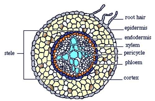
Internal structure of root
Uptake of water and salts:
Root hairs provide large surface area for absorption. They grow out into the spaces between soil particles where they are in direct contact with water. The cytoplasm of root hairs has a higher concentration of salts than soil water, so water moves by osmosis into root hairs. Salts also enter root hairs by diffusion or active transport. After their entry into root hairs, water and salts travel through intercellular spaces or through cells (via channels, called plasmodesmata) and reach xylem tissue. Once in xylem, water and salt are carried to all the aerial parts of the plant.
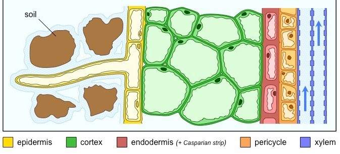
Uptake of water and salts ions by root
Q.2) Define transpiration and relate it with the cell surface and with stomatal opening and closing.
Answer:
Transpiration:
“Transpiration is the loss of water from the plant surface through evaporation.”
Sites of transpiration:
Loss of water may occur through stomata in leaves, through the cuticle present on leaf epidermis, and through special openings called lenticels present in the stems of some plants.
Stomatal transpiration:
“Most of the transpiration occurs through stomata and is called stomatal transpiration.”
Role of leaf surface area with transpiration:
A plant with a large surface area will transpire more water. The rate of transpirational water loss is greater per unit area of leaf surface. The mesophyll cells of the leaf provide a large surface area for the evaporation of water. Water is drawn from xylem to mesophyll cells, from where it comes out and makes a water-film on the cell walls of mesophyll. From here, water evaporates into the air spaces of the leaf. Water vapors then diffuse from air spaces towards stomata and then pass to outside air.

Events of transpiration in the section of a leaf
Opening and closing of stomata:
Most plants keep their stomata open during the day and close them at night.
Role of stomata in transpiration:
It is the responsibility of stomata to regulate transpiration via the actions of guard cells. The two guard cells of a stoma are attached to each other at their ends. The inner concave sides of guard cells that enclose a stoma are thicker than the outer convex sides. When guard cells get water and become turgid, their shapes are like two beans and the stoma between them opens. When guard cells lose water and become flaccid, their inner sides touch each other and stoma closes.
Concentration of solutes in guard cells:
The concentration of solutes (glucose) in guard cells is responsible for the opening and closing of stomata. Recent studies have revealed that light causes the movement of potassium ions from epidermal cells into guard cells. Water follows these ions and enters guard cells. Thus their turgidity increases and stoma opens. As the day progresses, guard cells make glucose i.e. become hypertonic. So water stays in them. At the end of the day, potassium ions flow back from guard cells to epidermal cells and the concentration of glucose also falls. Due to it, water moves to epidermal cells and guard cells lose turgor. It causes a stomach ache.
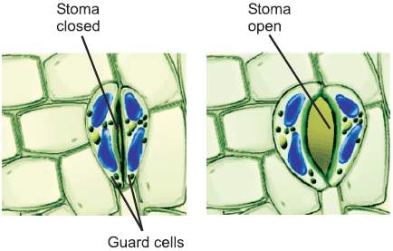
Opening and closing of stomata
Q.3) How do different factors affect the rate of transpiration?
Answer:
Factors affecting the rate of transpiration:
The rate of transpiration is directly controlled by the opening and closing of stomata and it is under the influence of light. In strong light, the rate of transpiration is very high as compared to dim light or no light. Other factors which affect the rate of transpiration are given below.
1- Temperature:
Higher temperature reduces the humidity of the surrounding air and also increases the kinetic energy of water molecules. In this way, it increases the rate of transpiration. The rate of transpiration doubles with every rise of 100oC in temperature. But, very high temperatures i.e. 40-45 oC cause closure of stomata, so transpiration stops and the plant does not lose the much-needed water.
2- Air humidity:
When air is dry, water vapor diffuses more quickly from the surface of mesophyll cells into leaf air spaces and then from air spaces to outside. This increases the rate of transpiration. In humid air, the rate of the diffusion of water vapors is reduced and the rate of transpiration is low.
3- Air movement:
Wind (air in motion) carries away the evaporated water from leaves and it causes an increase in the rate of evaporation from the surfaces of the mesophyll. When the air is still, the rate of transpiration is reduced.
4- Leaf surface area:
The rate of transpiration also depends upon the surface area of the leaf. More surface area provides more stomata and there is more transpiration.
Q.4) is transpiration a necessary evil? Give comments.
Answer:
Transpiration a necessary evil:
Transpiration is called a necessary evil. It means that transpiration is a potentially harmful process but is unavoidable too. Transpiration may be a harmful process in the sense that during the conditions of drought, loss of water from plants results in serious desiccation, wilting and often death.
On the other hand, transpiration is necessary too. It creates a pulling force called transpirational pull which is principally responsible for the conduction of water and salts from roots to the aerial parts of the plant body. When water transpired from the surfaces of plants, it left a cooling effect on plants. This is especially important in warmer environments. Moreover, the wet surfaces of leaf cells allow gaseous exchange.
Q.5) Explain the movement of water in terms of transpirational pull.
Answer:
Transpiration Pull:
“Transpiration pull is the force which aids in drawing the water upward from roots to leaves.”
When a leaf transpires (loses water), the water concentration of its mesophyll cells drops. This drop causes water to move by osmosis from the xylem of the leaf into mesophyll cells. When one water molecule moves up in the xylem of the leaf, it creates a pulling force that continues all the way to root. This pulling force created by the transpiration of water is called transpiration pull. It also causes water to move transversely (from root epidermis to cortex and pericycle).
Reasons for transpiration pull:
Following are the reasons for the creation of transpiration pull.
- Water is held in a tube (xylem) that has a small diameter.
- Water molecules adhere to the walls of the xylem tube (adhesion).
- Water molecules cohere to each other (cohesion).
These attractions make an overall tension among water molecules. This tension forms columns of water. The columns of water move from root to shoot and the water content of the soil enters in these ‘columns’.

Transport of Water
Q.6) Describe the theory of pressure flow mechanism to explain the translocation of food in plants.
Answer:
Pressure-flow mechanism:
“The mechanism by which sugars are transported through the phloem, from sources to sinks, is called pressure flow. At the sources (leaves), sugar molecules are moved into the sieve elements (phloem cells) through active transport.”
Translocation of food through phloem:
Phloem is responsible for transporting food substances throughout the plant body. The glucose formed during photosynthesis in mesophyll cells, is used in respiration and the excess of it is converted into sucrose. In most plants, food is transported in the form of sucrose. The currently accepted hypothesis states that transport of food is through a pressure-flow mechanism. In a pressure-flow mechanism, food is moved from sources to sinks.

Transport of Food
i- Sources (leaf or storage organs):
Sources include the exporting organs, typically a mature leaf or storage organ.
ii- Sinks:
Sinks are the areas of active metabolism or storage e.g. roots, tubers, developing fruits and leaves, and growing regions. A storage organ is capable of storing food and exporting the stored materials.
Example:
The root of beet is a sink in the first growing season, but becomes a source in the next growing season, when sugars are utilized in the growth of new shoots.
Mechanism of food transport:
At source, food (sugars) is moved by active transport into the sieve tubes of phloem. Due to the presence of sugar in sieve tubes, their solute concentration increases and water enters them from xylem (via osmosis). This results in higher pressure of water in these tubes, which drives the solution of food towards the sink.
At the sink end, food is unloaded by active transport. Water also exits from the sieve tubes. The exit of water decreases pressure in sieve tubes, which causes a mass flow from the higher pressure at the source to the lowered pressure at the sink.
Q.7) List the functions of the components of blood.
Answer:
Blood:
Blood is a specialized body fluid (a connective tissue) that is composed of a liquid called blood plasma and blood cells.
The weight of blood in our body is about 1/12th of our body. The average adult body has about 5 liters of blood.
Components of blood and their functions:
1- Blood Plasma:
Plasma is primarily water in which proteins, salts, metabolites, and wastes are dissolved.
Composition of blood plasma:
Water constitutes about 90-92% of plasma and 8-10% are dissolved substances. Salts make up 0.9 % of plasma, by weight. Sodium chloride (the table salt) and salts of bicarbonate are present in considerable amounts. Ca, Mg, Cu, K and Zn are found in trace amounts. Changes in the concentration of any salt can change the pH of blood (normal is 7.4). Proteins make 7-9 % by weight of plasma. The important proteins present in plasma are antibodies, fibrinogen (a blood clotting protein), albumin (maintains the water balance of blood) etc. Plasma also contains the digested food (absorbed from the digestive system), nitrogenous wastes and hormones. Respiratory gasses i.e. CO2 and O2are present in the plasma.
Blood cells and cell-like bodies:
These include red blood cells (erythrocytes), white blood cells (leukocytes) and platelets (thrombocytes).
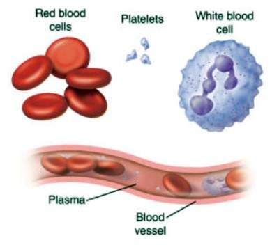
Different cells and cell like bodies in blood
1- Red blood cells (RBCs):
These are the most numerous of blood cells. A cubic millimeter of blood contains 5 to 5.5 million of RBCs in males, and 4 to 4.5 million in females. When RBCs are formed, they have a nucleus. In mammals, when a red blood cell matures, its nucleus is lost. After the loss of the nucleus, RBC enters the blood.
Composition of RBCs:
About 95% of the cytoplasm of RBCs is filled with hemoglobin, which transports O2 and small amounts of CO2. The remaining 5% consists of enzymes, salts and other proteins. RBCs are biconcave and have an elastic cell membrane. In the embryonic and fetal life, they are formed in the liver and spleen. In adults, they are formed in the red bone marrow of short and flat bones, such as sternum, ribs, and vertebrae.
Lifespan of RBCs:
An average life span of RBC is about four months (120 days) after which it breaks down in the liver and spleen by phagocytosis.
2- White Blood Cells/Leukocytes (WBCs):
These are colorless, because they do not contain pigments. They are not confined to blood vessels and also migrate out into tissue fluid. One cubic millimeter of blood contains 7000 to 8000 WBCs.
Lifespan of WBCs:
Their lifespan ranges from months to even years, depending on the body’s needs. WBCs function as the main agents in the body’s defense system.
Types of WBCs:
There are two main types of WBCs.
i- Granulocytes:
Granulocytes have granular cytoplasm. These include neutrophils (destroy small particles by phagocytosis), eosinophils (break inflammatory substances and kill parasites) and basophils (prevent blood clotting).
ii- Agranulocytes:
Agranulocytes have clear cytoplasm and include monocytes (produce macrophages which engulf germs) and B and T lymphocytes (produce antibodies and kill germs).
3- Platelets (Thrombocytes):
They are not cells but are fragments of large cells of bone marrow, called megakaryocytes. They do not have any nucleus and any pigment. One cubic millimeter of blood contains 250,000 platelets.
Lifespan of thrombocytes:
The average lifespan of a blood platelet is about 7 to 8 days.
Function of platelets:
Platelets help in blood clotting. The clot serves as a temporary seal at the damaged area.
Q.8) How do we classify blood groups in terms of the ABO and the Rh blood group systems ?
Answer:
Blood group systems:
Blood group systems are a classification of blood; based on the presence or absence of antigens on the surface of red blood cells.
Antigen:
“An antigen is a molecule that can stimulate an immune response (antibody production etc).”
1- ABO Blood Group System:
ABO is the most important blood group system in humans. It was discovered by the Austrian scientist Karl Landsteiner, who found four different blood groups (blood types) in 1900. He was awarded the Nobel Prize in Medicine for his work.
Types of blood groups:
In this system, there are four different blood groups which are distinct from each other on the basis of specific antigens (antigen A and B) present on the surface of RBCs.
- A person having antigen A has blood group A
- A person having antigen B has blood group B
- A person having both antigens has blood group AB
- A person having none of the A and B antigens has blood group O.
Antibodies in blood serum:
After birth, two types of antibodies i.e. anti-A & anti-B antibodies appear in the blood serum of individuals. These antibodies are present according to the absence of the corresponding antigen.
- In persons with blood group A, antigen A is present and antigen B is absent. So their blood will contain anti-B antibodies.
- In persons with blood group B, antigen B is present and antigen A is absent. So their blood will contain anti-A antibodies.
- In persons with blood group AB, antigens A & B are present i.e. neither is absent. So their blood serum will contain no antibody.
- In persons with blood group O, neither antigen A nor antigen B is present i.e. both are absent. So their blood serum will contain both antibodies i.e. anti-A & anti-B.
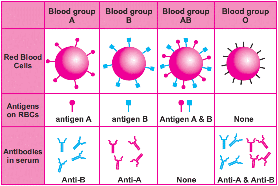
ABO Blood group system showing presence and absence of antigens and antibodies
2- Rh Blood Group System (+ ve & – ve blood group system):
In the 1930s, Karl Landsteiner discovered the Rh-blood group system.
Types of blood group in Rh- system:
In this system, there are two blood groups i.e. Rh-positive and Rh-negative. These blood groups are distinct from each other on the basis of antigens called Rh factors (first discovered in Rhesus monkeys), present on the surface of RBCs.
- A person having Rh factors has blood group Rh-positive
- A person not having Rh factors has blood group Rh-negative.
Unlike the naturally occurring anti-A & anti-B antibodies of the ABO-system, an Rh-negative person does not produce anti-Rh antibodies unless Rh-factor enters in his / her blood.
Q.9) State the signs and symptoms, causes and treatments of leukemia and thalassemia.
Answer:
1- Leukemia (blood cancer):
Leukemia is the production of a great number of immature and abnormal white blood cells.
Causes:
This is caused by a cancerous mutation (change in a gene) in bone marrow or lymph tissue cells. The mutation results in uncontrolled production of defective white blood cells (leukocytes).
Treatment:
It is a very serious disorder and patients need to change blood regularly with normal blood, obtained from donors. It can be cured by bone marrow transplant. It is effective in most cases but very expensive.
2- Thalassaemia:
It is also called Cooley’s anemia in the name of Thomas B. Cooley, an American physician.
Cause:
It is a genetic problem due to mutations in the gene of hemoglobin. The mutation results in the production of defective hemoglobin and the patient cannot transport oxygen properly.
Treatment:
The blood of these patients is to be replaced regularly, with normal blood. It can be cured by bone marrow transplant but it does not give a 100% cure rate.
Q.10) What four chambers make the human heart and how blood flows through these chambers ?
Answer:
Human heart:
“Heart is a muscular organ responsible for pumping blood through blood vessels by repeated contractions.”
Cardiac muscles:
The bulk of the walls of heart chambers is made of cardiac muscles.
Location of heart:
In the human body, the heart is situated between the lungs, in the middle of the chest cavity (thorax) under the breastbone.
Pericardium:
The heart is enclosed in a sac known as the pericardium. There is a fluid, known as pericardial fluid, between the pericardium and heart walls. It reduces friction between the pericardium and heart, during heart contractions.
The internal structure of the heart:
The human heart consists of four chambers, like the hearts of birds and other mammals. The upper thin-walled chambers are called left and right atria (singular ‘atrium’), and the lower thick-walled chambers are called left and right ventricles. The left ventricle is the largest and strongest chamber in the heart.
Heart double pump mechanism:
The human heart works as a double pump. It receives deoxygenated (with less oxygen) blood from the body and pumps it to the lungs. At the same time, it receives oxygenated (with more oxygen) blood from the lungs and pumps it to the whole body. Inside heart chambers, the deoxygenated and oxygenated blood are kept separated.
Pathway of deoxygenated blood in heart:
- The right atrium receives deoxygenated blood from the body via the main veins i.e. superior and inferior vena cavae.
- When the right atrium contracts it passes the deoxygenated blood to the right ventricle.
- The opening between the right atrium and right ventricle is guarded by a valve known as the tricuspid valve (because it has 3 flaps).
- When the right ventricle contracts, the blood is passed to the pulmonary trunk, which carries blood to the lungs.
- The tricuspid valve prevents the backflow of blood from the right ventricle to the right atrium.
- At the base of the pulmonary trunk, the pulmonary semilunar valve is present which prevents the backflow of blood from the pulmonary trunk to the right ventricle.
Pathway of oxygenated blood in the heart:
- The oxygenated blood from the lungs is brought by pulmonary veins to the left atrium
- Left atrium contracts and pumps this blood to the left ventricle.
- The opening between the left atrium and left ventricle is guarded by a valve known as a bicuspid valve (because it has two flaps).
- When the left ventricle contracts, it pumps the oxygenated blood into the aorta, which carries blood to all parts of the body (except the lungs).
- The bicuspid valve prevents the backflow of blood from left ventricle to the left atrium.
- At the base of the aorta, an aortic semilunar valve is present which prevents the backflow of blood from the aorta to the left ventricle.

Human heart; Structure and blood flow
Q.11) Compare the structure and function of an artery, a vein and a capillary.
Answer:
| Comparison of arteries, capillaries, and veins | |||
| Characteristics | Arteries | Capillaries | Veins |
| Function | Arteries carry blood away from the heart | Capillaries allow the exchange of materials between blood and tissues | Veins carry blood towards the heart |
| Thickness and elasticity in walls | They are thick and elastic | One cell thick and non-elastic walls | They are thin and less elastic |
| Muscles in walls | Thick | No muscles present | Thin muscle |
| Blood pressure | High Bp | Medium Bp | Low Bp |
| Valves | No valves | No valves | Valves present |
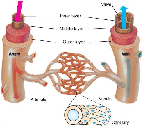
Blood vessels; Artery, Capillary, and Vein
Q.12) Draw diagrams that can illustrate the origins, locations, and target areas of the main arteries in the human blood circulatory system.
Answer:
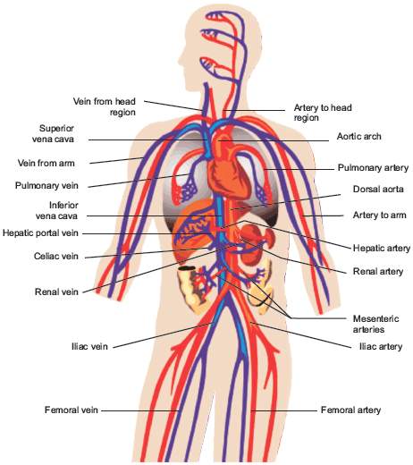
Human Arterial system (Red color in the diagram is the artery)
Q.13) Draw diagrams that can illustrate the areas and locations of the main veins in the human blood circulatory system.
Answer:
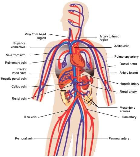
Venous system in Human body (Blue color shows veins)
Q.14) How would you differentiate between atherosclerosis and arteriosclerosis?
Answer:
Atherosclerosis and Arteriosclerosis:
Atherosclerosis and arteriosclerosis are the diseases of arteries. These diseases also lead to heart diseases.
Atherosclerosis:
“Atherosclerosis is a chronic disease in which narrowing of arteries occurs due to the accumulation of fatty materials, cholesterol, or fibrin in arteries.”
When this condition is severe, arteries can no longer expand and contract properly, and blood moves through them with difficulty.
Reason of atherosclerosis:
Accumulation of cholesterol is the prime contributor to atherosclerosis. It results in the formation of multiple deposits called plaques within arteries. Plaques can form blood clots called thrombi within arteries. If a thrombus dislodges and becomes free-floating, it is called an embolus.
Arteriosclerosis:
Arteriosclerosis is a chronic disease in which the hardening of arteries occurs when calcium is deposited in the walls of arteries.
Reason:
It can happen when atherosclerosis is severe.
Q.15) State the causes, treatments, and prevention of myocardial infarction
Answer:
Myocardial infarction:
“Myocardial infarction (heart attack)occurs when blood flow decreases or stops to a part of the heart, causing damage to the heart muscle.”
The term myocardial infarction is derived from myocardium (the heart muscle) and infarction (tissue death).
Reason for heart attack:
A heart attack occurs when the blood supply to a part of the heart is interrupted and leads to the death of heart muscles. A heart attack may be caused by a blood clot in coronary arteries. It is a medical emergency and the leading cause of death for both men and women all over the world.
Symptoms:
Severe chest pain is the most common symptom of myocardial infarction and maybe in the form of the sensation of tightness, pressure, or squeezing. Pain radiates most often to the left arm, but may also radiate to the lower jaw, neck, right arm, and back. Loss of consciousness and even sudden death can occur in myocardial infarction.
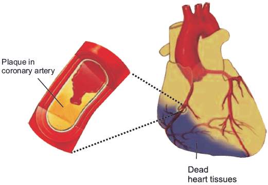
Atherosclerosis and resulting myocardial infarction
Treatment:
Immediate treatment for suspected acute myocardial infarction includes oxygen supply, aspirin, and a sublingual tablet of glyceryl trinitrate. Most cases of myocardial infarction are treated with angioplasty (mechanical widening of a narrowed or totally obstructed blood vessel) or bypass surgery (surgery in which arteries or veins from elsewhere in the patient’s body are grafted to the coronary arteries to improve blood supply to heart muscles).
Short Questions
Q.1) What are lenticels and where are the; found in the plant body ?
Answer:
Lenticels:
“A lenticel is one of many raised pores in the stem of a woody plant that allows gas exchange between the atmosphere and the internal tissues.”
Location of lenticels:
A lenticel is an airy aggregation of cells within the structural surfaces of the stems, roots, and other parts of vascular plants.
Function of lenticels:
It functions as a pore, providing a medium for the direct exchange of gasses between the internal tissues and atmosphere.
Q.2) What is the role of potassium ions in the opening of stomata ?
Answer:
Role of potassium in the opening of stomata:
Light causes the movement of potassium ions from epidermal cells into the guard cells. Water follows these ions and enters the guard cells, as a result, guard cells become turgid and stomata open.
As the day progresses, guard cells make glucose i.e. become hypertonic so water stays in them. At the end of the day, potassium ions flow back from the guard cells toward the epidermal cells and the concentration of glucose also falls. Due to this, water moves to epidermal cells and guard cells lose turgor, as a result, stomata close.
Q.3) Define the cohesion-tension theory.
Answer:
Cohesion-Tension Theory:
“The force by which water is raised to considerable heights in plants is termed as a cohesion-tension theory.”
“Or”
“The cohesion-tension theory is a theory of intermolecular attraction that explains the process of water flow upwards (against the force of gravity) through the xylem of plants.”
- According to this theory, the mechanism by which water (along with dissolved materials) is carried upward through the xylem is the transpirational pull.
- Transpiration creates a pressure difference that pulls water and salts up from their roots.
Q.4) What do you mean by sources and sinks according to the pressure flow mechanism ?
Answer:
Source:
Sugar sources are plant organs such as leaves that produce sugars.
Sinks:
Sugar sinks are plant organs such as roots, tubers (underground stems), and bulbs (swollen leaves) that consume or store sugars.
Q.5) What are the two main types of white blood cells? How do they differ?
Answer:
Types of WBCs:
There are two main types of WBCs.
i- Granulocytes
ii- Agranulocytes
Difference between granulocytes and agranulocytes:
| Granulocytes | Agranulocytes |
| Granulocytes have granular cytoplasm. | Agranulocytes have clear cytoplasm |
| These include neutrophils (destroy small particles by phagocytosis), eosinophils (break inflammatory substances and kill parasites) and basophils (prevent blood clotting). | These include monocytes (produce macrophages which engulf germs) and B and T lymphocytes (produce antibodies and kill germs). |
| Granulocytes are originated from the bone marrow of the human beings | Agranulocytes originate from the lymphoid. |
| These are polymorphonuclear leukocytes containing two to four lobes | These are mononuclear leukocytes containing one lobe |
Q.6) You see pus at the site of infection on your skin. How is it formed?
Answer:
Pus:
White blood cells die in the process of fighting against foreign particles. These dead blood cells accumulate and make a white substance called pus, seen at the infection sites.
Process of pus formation:
Pus is produced from the dead and living cells that travel into intercellular spaces around the affected cells. The most common agents that induce pus formation are bacteria, such as Staphylococcus aureus. In addition, some chemical agents can also cause pus creation.
Q.7) What role does the pericardial fluid play?
Answer:
Pericardial fluid:
The pericardial fluid reduces friction within the pericardium by lubricating the epicardial surface allowing the membranes to glide over each other with each heartbeat.
Q.8) Define the terms systole and diastole.
Answer:
Systole:
The time at which ventricular contraction occurs is called systole. Systole is the contraction of the chambers of the heart, driving blood out of chambers.
Diastole:
Diastole is the period of time when the heart fills with blood after systole (contraction). Ventricular systole is the period during which the ventricles are relaxing, while atrial diastole is the period during which the atria are relaxing.
Science Technology And Society
Q.1) State vascular surgery as one of the major fields in the careers.
Answer:
Vascular surgery:
“Vascular surgery is a surgical field in which diseases of the vascular system, or arteries, veins, and lymphatic circulation, are managed by medical therapy and surgical methods.”
Many patients referred to a vascular specialist do not require surgical or radiological intervention, but rather reassurance and lifestyle advice (lose weight, take regular exercise) coupled with measures to reduce their future risk of heart disease and stroke (antiplatelet and lipid-lowering therapy, blood pressure control). Additionally, smoking is a major cause of vascular disease and over 80% of vascular patients are current or ex-smokers.
Patients requiring vascular surgery suffer from many different vascular disorders that adversely affect the quality of life. The core activities of the vascular specialist include:
- Promoting cardiovascular health
- Improving the quality of life in patients with vascular disease
- Assisting colleagues from other specialties with the control of vascular bleeding
- Assisting colleagues in the management of the vascular complications of diabetes and renal disease
- Providing a renal access service for patients
Q.2) Identify that cardiovascular disorders are the major cause of sudden non-accidental deaths.
Answer:
Sudden cardiac death (SCD):
Sudden cardiac death (SCD) is a sudden, unexpected death caused by a change in heart rhythm (sudden cardiac arrest). It is the largest cause of natural death in the U.S., and all over the world. SCD is responsible for half of all heart disease deaths.
Causes of sudden cardiac death:
Sudden cardiac arrest is different from a heart attack, which occurs when blood flow to a portion of the heart is blocked. However, a heart attack can sometimes trigger an electrical disturbance that leads to sudden cardiac arrest. Other cardiovascular diseases can also lead to sudden cardiac arrest. Some of them are:
1- Coronary artery disease:
Most cases of sudden cardiac arrest occur in people who have coronary artery disease. In coronary artery disease, your arteries become clogged with cholesterol and other deposits, reducing blood flow to your heart. This can make it harder for your heart to conduct electrical impulses smoothly.
2- Heart attack:
If a heart attack occurs, often as a result of severe coronary artery disease, it can trigger ventricular fibrillation and sudden cardiac arrest. In addition, a heart attack can leave behind areas of scar tissue. Electrical short circuits around the scar tissue can lead to abnormalities in your heart rhythm.
3- Enlarged heart (cardiomyopathy):
This occurs primarily when your heart’s muscular walls stretch and enlarge or thicken. In both cases, your heart’s muscle is abnormal, a condition that often leads to heart tissue damage.
4- Electrical problems in the heart:
In some people, the problem is in the heart’s electrical system itself instead of a problem with the heart muscle or valves. These are called primary heart rhythm abnormalities.
Q.3) Explain the social as well as personal factors that contribute to. cardiovascular disorders in Pakistan.
Answer:
Cardiovascular diseases (CVD):
Cardiovascular diseases (CVD) are one of the commonest causes of disease and death not only in the developed but also in the developing world. The common risk factors are like smoking, diabetes, high cholesterol diet, lack of physical activity, hypertension and obesity.
1- Tobacco Consumption:
The relationship between tobacco use and heart attack is well established. Cigarette smoking increases the risk of CVD death by 70 % compared with not smoking.
2- Hypertension:
High levels of systolic and diastolic blood pressures have a strong relationship with heart attack.
3- Obesity:
Many of the lifestyle factors associated with overweight and obesity are found in urban areas. The main causes are increased consumption of energy-dense foods high in saturated fats and sugars and reduced physical activity.
4- Sedentary lifestyle:
A sedentary lifestyle is a type of lifestyle with little or no physical activity. Sedentary behavior includes reading, computer use, watching television, office work, and cell phone use. Such a lifestyle leads to diabetes and heart problems due to obesity.


